Digital Dental Radiographs
Benefits of Dental Xrays
Dental x-rays are vital tools to determine the proper diagnosis and treatment for many diseases and traumatic injuries. They help us to detect problems that would be missed by only looking directly at your mouth.
Problems that radiographs detect:
- Cavities between teeth or under fillings
- Delayed or altered teeth and jaw development in children and teens
- Bone loss from severe gum disease
- Infections at tips of dental roots
As helpful and radiographs are we try to limit them to only the necessary films as they require low dose ionizing radiation. Therefore, we always follow the ALARA principle when prescribing radiographs.
ALARA: As Low As Resonably Achievable
The American Dental Association (ADA) is confident that x-rays are safe except in cases of pregnancy. They recommend the ALARA principle (as low as reasonably achievable) is used for best results. they only should be performed when an appropriate healthcare professional has determined they would provide critical information. Therefore, the very first level of patient protection is minimizing radiation to ALARA.
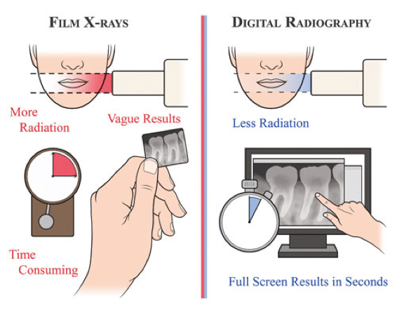
We take digital radiographs as they require 90% less radiation dose than conventional E speed film radiographs (Jayachandran 2017) and provide nearly instantaneous images without chemicals.

To help limit the amount of radiation exposure, we use full lead aprons with a thyroid collar.
Are Dental Xrays Safe?
Dental radiographs are safe when used reasonably. In fact they are so safe that the American College of Obstetricians and Gynaecologist evaluated the data and found dental xrays safe at any stage of pregnancy. The low dose of our dental radiograph systems were found to have no significant effect on unborn children and instead delaying dental treatment, including xrays, could lead to more serious problems like an infection.
We do not take radiographs on pregnant patients in the absence of acute issues, but isn’t it comforting to know that they are safe enough even for pregnant women and their unborn children.
We always try to minimize radiographs to only when necessary and are proud that we have selected systems that deliver quality images at the lowest reasonable dose.
Common Types of Dental Xrays
There are several types of dental rays. Each one helps the team see different areas of you mouth.
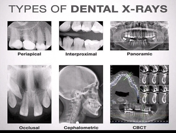
Common xrays used in the office include bite-wings, periapicals, cephalometric, CBCT and panoramic rays.

Periapicals allow us to observe conditions below the gumline as they show the bone and roots of teeth. They can be taken of front or back teeth. Panoramic radiographs are taken on a machine that rotates around the head to produce a long film that shows the entire jaw, joint, and sinuses in one image. Cone-beam computed technology (CBCT) images are taken in series to create a three-dimensional image. Because it relies on multiple images, radiation exposure is higher for CBCT surveys than that of the other xrays. CBCT is reserved for use when more detailed anatomical information is needed.
Xray frequency and types
The type, number and frequency of radiographs are prescribed by your dentist based on multiple factors such as:
- Your current oral health
- Your age
- Your risk for tooth decay and gum disease.
- Date of last xrays taken
Conclusion
Dental radiographs are a safe way for us to observe conditions beyond the surfaces of your gums, teeth, cheeks, and tongue. They help us to spot decay, keep an eye on advanced gum disease, and monitor tooth development in children. If you ever have any questions or concerns, we welcome your questions and would love to have you call us at (408)523-4030 or email dr@jenchiangdds.com. Our goals is to educate and inform our patients so that they are able to take on a proactive in your healthcare decisions.
